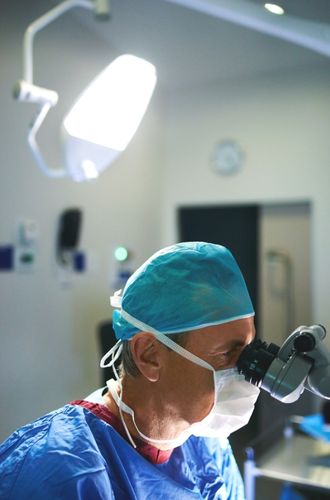
Dr. Nilesh Satbhai has years of experience performing microsurgery procedures and uses only the latest techniques and technology. Dr. Nilesh offers a wide range of services, from tissue transfer to composite tissue transplantation, to meet your needs.
Microsurgery is a mechanism used by many plastic surgeons to perform specific procedures, including tissue transfer from one part of the body to another (free tissue transfer), reattachment of several parts (replantation), and composite tissue transplantation.
Microsurgery is a surgical procedure that combines magnification with advanced diploscopes, specialized precision tools, and various other operating techniques. These techniques are mainly used to anastomose small blood vessels (arteries and veins) and to close the nerves together.
Microsurgery is indicated when the surgical procedure to be performed is too delicate or complex to be carried out by conventional surgical means.
Various procedures involved in microsurgery are:
An arteriovenous fistula (AVF) is not a normal connection between an artery and a vein. Sometimes, arteriovenous fistula is present at birth (congenital), or it may develop after birth, and sometimes they are the result of an injury (acquired).
The most common indication for the creation of an arteriovenous (AV) fistula is renal failure which requires chronic hemodialysis. It is performed to create a native fistula, although prosthetic material may be needed if a suitable vein is not available.
Some of the common forms of arteriovenous fistulas are:
Arteriovenous fistulas can be treated with endovascular embolization, microsurgery, or stereotactic radiosurgery.
Endovascular embolization: The most common form of treatment for an arteriovenous fistula. EE procedure is performed by placing a catheter into an artery (usually the femoral artery in the front of the hip). Then, with the help of fluoroscopic or X-ray imaging, your surgeon will move it to the location of the fistula. Once the connection between the artery and the vein is closed, the AVF is cured and usually does not reoccur.
Microsurgery: Microsurgery is the most suitable treatment for a dural, brain, or spinal AVF, either alone or in combination with endovascular embolization. Dr. Nilesh Satbhai can tell you before the treatment if microsurgery will be necessary. With microsurgery, neurosurgery is also performed to visualize the AVF under a microscope and Dr. Nilesh Satbhai will place a titanium clip over the abnormal connection to prevent blood from flowing abnormally from the artery to the vein. During the procedure, Dr. Nilesh Satbhai can see the abnormal blood flow stop, and the abnormal vein changes from red (when it is carrying arterial blood) to blue, which is the normal color of veins. Immediately after the procedure, Dr. Nilesh Satbhai usually performs an angiogram to confirm that the AVF has been completely treated.
Following are a few of the most important benefits of this treatment:
As with any surgery, there are certain risks associated with AV fistula management. However, these risks are usually rare and can be effectively managed by Dr. Nilesh Satbhai . Some of the most common risks of AV fistula management include:
Local flaps are those that are derived from the immediate area of resection, that is, local flaps are the use of tissue adjacent to the defect.
Common examples of these include:
These types of flaps are advanced, transposed, or rotated into position and supplied by either an axial pattern or a random pattern.
Regional flaps are defined as those that are located near a defect but are not in immediate proximity. Regional flaps are frequently harvested from the neck, chest, or axilla.
Common examples of these include:
Although all surgeries come with risks, those associated with local and regional flaps are relatively rare. Dr. Nilesh Satbhai is skilled in managing these risks so that patients can feel reassured about their surgery. Some of the most common potential complications include:
Perforator flap surgery is a surgical technique used in reconstructive surgery where subcutaneous tissue or skin is removed from an adjacent or distant part of your body to reconstruct the part which is excised.
The vessels that supply blood to the flap are isolated perforators which are derived from a deep vascular system through your underlying muscle or intermuscular septa. Some of the perforators can have a mixed septal and intramuscular course before reaching the skin.
Septal perforator: A perforator that only traverses through the septum to supply the underlying skin is called a septal perforator.
Muscle perforator: A flap that is vascularised by a perforator traversing only through muscle to supply the underlying skin is called a muscle perforator.
Based on their vascular supply, these can be classified into direct and indirect perforators.
Direct perforators: Direct perforators pierce the deep fascia, they don’t traverse any other structural tissue.
Indirect perforators: Indirect perforators, before piercing the deep fascia, first run through the other structures.
Free perforator flaps A free flap is defined as a mass of tissue that has been taken away from the original site to be used in tissue transplantation. When your surgeon uses a free flap, the blood supply is cut and the pedicle reattached to the recipient’s vessels, performing a microsurgical anastomosis.
Pedicled perforator flaps Pedicled perforator flaps are transferred either by translation (also called “advancement”) technique or rotation technique.
Perforator flaps come with a small risk of complications. Dr. Nilesh Satbhai is an expert in mitigating these risks so that patients may rest easy during their surgery. The following are some of the potential risks of perforator flap:
The following procedures can be done for bone and soft tissue reconstruction after trauma.
The most commonly used tissue flaps for reconstruction are:
Fasciocutaneous flaps The anterolateral thigh (ALT) flap is the most popular flap for many reasons. The ALT provides extensive coverage, making it ideal for most defects.
Muscle flaps The latissimus dorsi (LD) flap, which is based on the largest muscle in the body, is used when a large amount of tissue and muscle is needed.
Bone fixation can be done by:
Fractures in cases of remarkable or persistent bone contamination should be treated with external fixation until the bone has healed fully.
During the first few days after surgery, it is normal to experience some pain and discomfort. Dr. Nilesh Satbhai will prescribe pain medication to help manage any discomfort. It is important to keep the surgical incisions clean and dry during this time. Dr. Nilesh Satbhai will provide specific instructions on how to care for your incisions.
The rehabilitation and recovery process after general microsurgery will vary depending on the individual case. It is important to follow Dr. Nilesh Satbhai ’s post-operative instructions carefully to ensure a smooth and successful recovery.
Most patients can return to work and their normal activities within a few weeks. However, it is important to avoid strenuous activity during the initial recovery period. Dr. Nilesh Satbhai will provide specific instructions on when you can resume your normal activities.
It is important to keep all follow-up appointments with Dr. Nilesh Satbhai . During these appointments, Dr. Nilesh Satbhai will monitor your recovery and ensure that you are healing properly.
The cost of general microsurgery in Mumbai will depend on the type of procedure you need, the location of the procedure, and whether or not you have insurance. During the consultation, Dr. Nilesh Satbhai will go over the expected costs with you.
Request Appointment
MBBS, MS, MCh
Dr. Nilesh Satbhai is a distinguished surgeon known for his meticulous approach, profound expertise, and compassionate care.
With a commitment to excellence, Dr. Nilesh Satbhai has redefined reconstructive and aesthetic surgery, setting new benchmarks in patient satisfaction and outcomes. His unique blend of artistry and science transforms not only physical appearances but also lives.
